Recently, Department of Pediatric Surgery of the First Affiliated Hospital (FAH) of Xi 'an Jiaotong University (XJTU) cooperated with Neonatal Ward, Department of Anesthesiology and Perioperative Medicine and Department of Radiology to successfully perform minimally invasive magnetic anastomosis under endoscope for a 3-day-old neonate with congenital esophageal atresia complicated with esophagotracheal fistula.
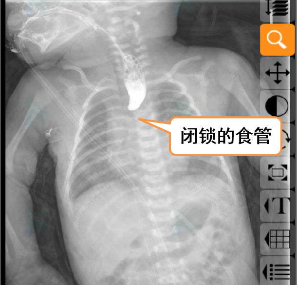
At 1 day after full-term delivery, physicians found that the esophagus was not connected with the stomach. Upon admission to the FAH, gastrointestinal radiography, chest CT scan and three-dimensional reconstruction of the airway detected tracheoesophageal fistula between the esophagus and trachea. The neonate was diagnosed with congenital esophageal atresia (type IIIA), i.e., congenital esophageal atresia complicated with esophagotracheal fistula, which should be treated immediately.
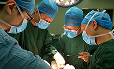
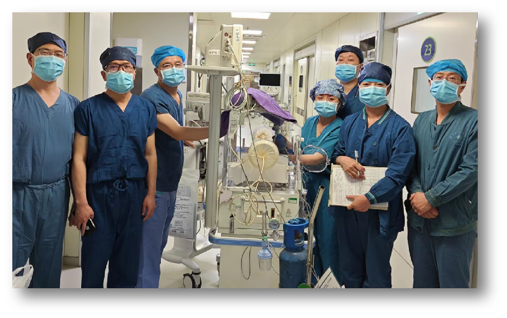
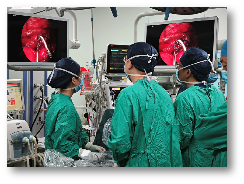
At present, the treatment of congenital esophageal atresia complicated with esophagotracheal fistula remains a global challenge. Traditional open surgery yields significant trauma, excessive bleeding, long recovery time and evident scar. However, it is highly demanding to simply perform thoracoscopic esophageal anastomosis. After multidisciplinary team (MDT) consultation and comprehensive evaluation of physician condition, and obtaining the consents of family members, Cao Zhenjie's team from Department of Pediatric Surgery eventually chose the world's cutting-edge minimally invasive magnetic anastomosis under 3-mm thoracoscope to repair esophageal atresia and esophagotracheal fistula. According to the structural characteristics of esophagus, the team specially invited Professor Liu Shiqi, a member of Professor Lyu Yi’s team and Director of Department of Neonatal Surgery in Xi 'an Children's Hospital, who accumulated abundant experience in the treatment of esophageal atresia using magnetic anastomosis, to design and manufacture a magnetic device suitable for the treatment of congenital digestive tract malformation in children. On May 13, the neonate was transferred into the operating room. Neonatal thoracoscopic surgery requires highly-demanding anesthesia skills during operation and requires one-lung ventilation. Wang Qiang, Director of Department of Anesthesiology and Perioperative Medicine delivered explicit anesthesia regimens and cooperated with Professor Li Xiaoquan and other physicians from Department of Neonatology to adopt electronic bronchoscope and visual laryngoscope for intraoperative visual monitoring to guarantee surgical success.
Surgical procedures must be carried out with significant cautions due to small chest volume, narrow surgical field, and adjacency between thoracic esophagus and the great vessels and critical nerves in infants. Intraoperatively, Professor Cao Zhenjie pinpointed the position of tracheoesophageal fistula. First, the proximal end of esophageal atresia was severed and the tracheoesophageal fistula was ligated under thoracoscope, and then the purse-string suture of unipolar magnets at both ends was carried out. Two magnets were inserted to connect the interrupted parts between the esophagus ends with magnetic stapler to restore the continuity of esophagus. During the operation, esophageal stricture at both ends was accurately closed, and the surgery was successfully implemented for the neonate.
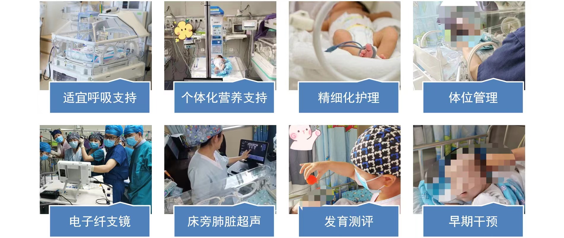
Postoperatively, the neonate was transferred to Neonatal Intensive Care Unit (NICU) for subsequent treatment. To guarantee the therapeutic effect of postoperative rehabilitation, Zhou Xihui's team determined an explicit rehabilitation plan and provided individualized daily nutritional support. The patient received alveolar lavage, bedside ultrasound dynamic monitoring, adjustment of anti-infection treatment regimen, maintenance of appropriate environmental temperature, maintenance of water and electrolyte balance in the body and early intervention and training delivered by rehabilitation physicians. After these interventions, physical condition was gradually stabilized and improved. Esophagography at postoperative 3 weeks showed that the anastomotic site was properly healed, no esophageal anastomotic leakage or stenosis, or recurrence of esophagotracheal fistula was observed. The esophageal morphology was properly restored.
The success of this procedure explores novel clinical treatment approaches for children with such complex diseases, and Department of Pediatric Surgery in the FAH becomes the pioneer to apply engineering medicine technique in the treatment of congenital digestive tract malformations.