In recent years, the incidence of endometrial cancer in China has continued to rise, with patients tending to be younger. Early diagnosis is key to improving survival rates and prognosis. Traditional pathological diagnosis based on endometrial cytology has long faced challenges such as difficult sampling, complex diagnosis, and a shortage of pathologists. China faces a shortage of tens of thousands of pathology professionals, and physicians capable of gynecological cytology diagnosis are even more scarce. These constraints have seriously limited the promotion of screening among high-risk populations at the primary care level and the improvement of diagnostic efficiency.
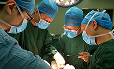
Cytological examination is minimally invasive and convenient for sampling, making it suitable for large-scale population screening and follow-up of high-risk groups. It is an indispensable part of the early diagnosis system for endometrial cancer. However, cytological diagnosis depends on physicians’ visual identification of subtle morphological changes in individual cells and cell clusters. Abnormal cells are distinguished based on features such as nuclear size, nuclear-cytoplasmic ratio, and chromatin distribution. The professional threshold is therefore extremely high, making it difficult to fully unlock its clinical value. In response to these clinical challenges, Chief Physician Li Qiling from the First Affiliated Hospital (FAH) of Xi’an Jiaotong University (XJTU) led a multidisciplinary team involving gynecologic oncology, cytopathology, and artificial intelligence. Through cross-disciplinary medical-engineering innovation, the team developed an artificial intelligence-based multiclass recognition-assisted diagnostic system for endometrial cytology. The project was successfully selected for the 2025 Artificial Intelligence Medical Device Innovation Task under the open competition mechanism.
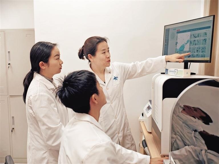
The system has achieved three core technological breakthroughs, establishing an intelligent closed loop covering the entire workflow from sample processing to report issuance.
First, the team built a domestically leading endometrial cytology image database. The database contains hundreds of thousands of high-resolution cell images, covering categories such as normal cells, benign lesions, atypical hyperplasia, and endometrial cancer. These images were cross-annotated through multiple rounds by senior pathology experts, laying a solid data foundation for deep learning of the artificial intelligence model.
Second, the team innovatively developed a deep learning model for multiclass cytological recognition. Breaking through the limitations of the traditional binary classification of benign and malignant lesions, the model incorporates an attention mechanism and a multitask learning framework. It can accurately capture dozens of quantitative cellular morphological features. The system can screen for suspicious malignant cells and precisely identify precancerous lesions, providing comprehensive and objective evidence for clinical decision-making.
Third, the team created a human-machine collaborative intelligent slide-reading workflow. Integrated with whole-slide digital scanning technology, artificial intelligence serves as a second reader by automatically marking and ranking suspicious fields of view. This frees physicians from the time-consuming task of searching through large numbers of images, allowing them to focus on professional judgment. At the same time, the system can also generate structured diagnostic reports, shorten slide-reading time, and reduce the risk of missed diagnosis caused by visual fatigue.
Currently, the project is accelerating medical device registration testing and clinical trials. As an important breakthrough for artificial intelligence in cytopathology, the system can also help address gaps in primary-level diagnosis and treatment, promote the flow of high-quality medical resources to the community, improve women’s access to gynecological screening services, and build a stronger safeguard for women’s health.