Recently, the Department of Radiation Oncology at the First Affiliated Hospital (FAH) of Xi’an Jiaotong University (XJTU) has successfully completed the first brachytherapy localization procedure in Northwest China using a radiotherapy-dedicated magnetic resonance imaging (MRI) simulator. This marks another significant breakthrough for the FAH in the field of image-guided tumor radiotherapy technologies.
As the first radiotherapy-dedicated MRI simulator introduced into clinical practice in Northwest China, the equipment provides higher-resolution soft tissue contrast during the treatment planning stage. In particular, for gynecological tumor brachytherapy, it allows clear visualization of tumor extent and the location of adjacent organs at risk, thereby offering precise imaging evidence for target delineation and dose optimization. Compared with conventional CT simulation, MRI simulation not only improves the accuracy of distinguishing between tumors and normal tissues, but also significantly reduces dose deviations caused by the limitations of imaging.
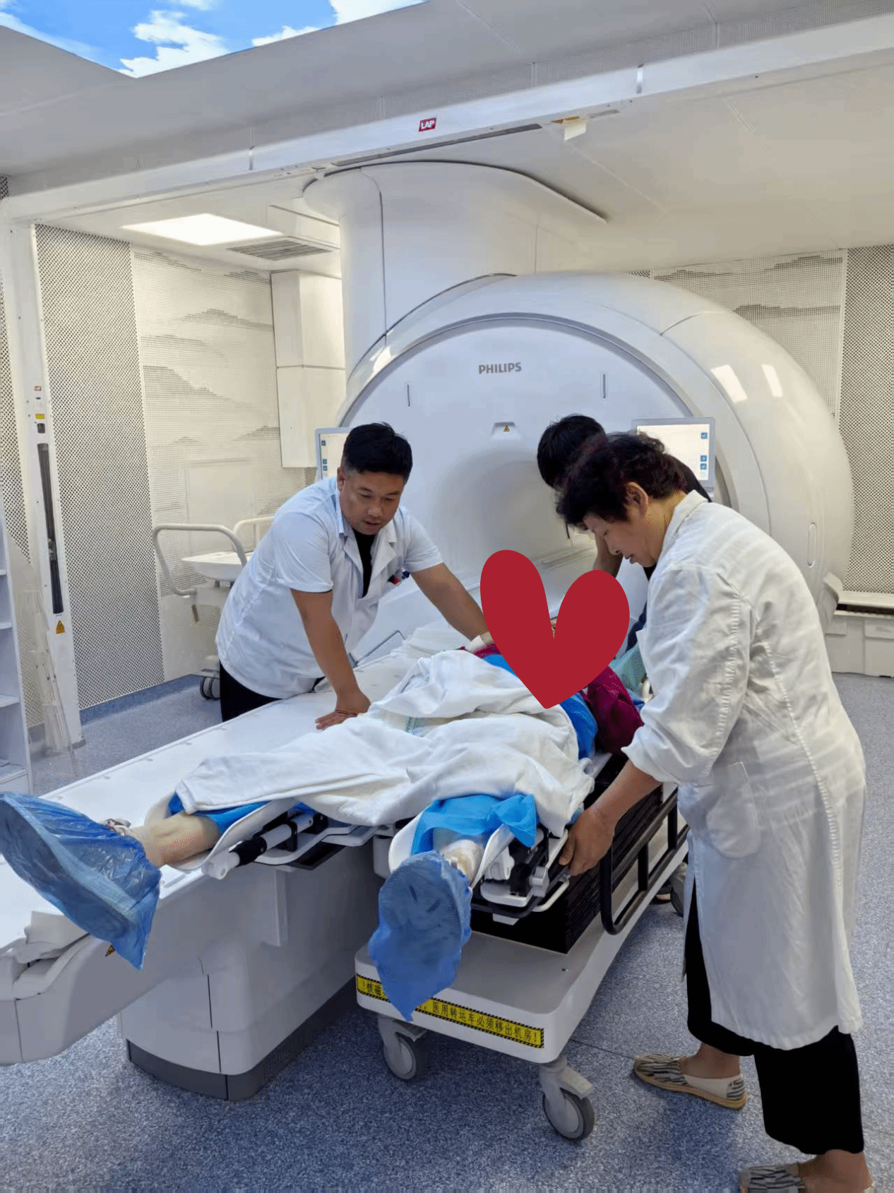
In this clinical application, under the leadership of Professor Zhang Xiaozhi, the team from the Department of Radiation Oncology collaborated closely with gynecologic oncologists, medical physicists, technologists, and nursing staff to successfully complete the MRI-based localization process for brachytherapy patients. Guided by high-precision imaging, the team further optimized individualized radiotherapy treatment planning, achieving dual improvements in both treatment accuracy and patient safety.