To enable early warning of cerebral palsy (CP), the leading cause of childhood disability, the First Affiliated Hospital (FAH) of Xi’an Jiaotong University (XJTU), in collaboration with more than ten medical institutions nationwide, has for the first time developed an intelligent risk prediction model for infants aged 6 months to 2 years based on routine magnetic resonance imaging (MRI), thereby addressing the clinical challenge of late diagnosis and delayed intervention. The related findings were published in eClinicalMedicine, a Lancet specialty journal, and in IEEE Trans Med Imaging, a leading journal in medical imaging, providing a “China solution” for the early prevention and control of CP worldwide.
CP ranks as the leading cause of disability among children in China, with white matter injury, occurring in more than 20% of preterm infants, as its primary cause. Conventional clinical diagnosis is often delayed until after the age of two, thereby missing the golden window of neuroplasticity for intervention. The FAH of XJTU, together with 12 medical institutions nationwide including the First Affiliated Hospital of Henan University of Chinese Medicine and the Affiliated Hospital of Zunyi Medical University, has for the first time achieved early and accurate prediction of CP based on routine MRI images commonly available in primary healthcare. The study was published in eClinicalMedicine, a Lancet specialty journal, with Dr. Huang Tingting as the first author and Prof. Yang Jian as the corresponding author.
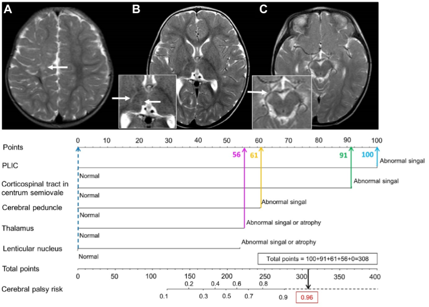
The research team identified abnormal signals or atrophy in the posterior limb of the internal capsule, the corticospinal tract at the centrum semiovale, the cerebral peduncle, the thalamus, and the lentiform nucleus as five independent predictive factors of CP. Based on these findings, they innovatively developed a visual nomogram model that translates complex medical imaging features into an intuitive risk score, with risk probabilities clearly displayed. The model demonstrated excellent performance in multicenter validation involving 383 infants. The external validation cohort achieved an AUC of 0.92 (95% CI: 0.86-0.97). Blind testing by 11 physicians with different levels of experience yielded an average AUC of 0.96, with sensitivity and specificity of 90% and 88%, respectively. The model requires only routine MRI without the need for costly advanced sequences, making it suitable for promotion in resource-limited regions.
Article link:
https://www.thelancet.com/journals/eclinm/article/PIIS2589-5370(25)00296-2/fulltext
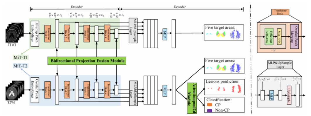
The team led by Prof. Yang Jian at the FAH of XJTU, together with Prof. Sun Jian’s team at the School of Mathematics and Statistics, further integrated artificial intelligence to achieve automated recognition of lesions in key brain regions and multimodal feature fusion, and preliminarily established an intelligent risk assessment system for CP. The related algorithms were published in IEEE Trans Med Imaging, with Dr. Qi Kai and Dr. Huang Tingting as co-first authors, and Prof. Sun Jian and Prof. Yang Jian as co-corresponding authors.
Article link:
https://ieeexplore.ieee.org/document/11018476