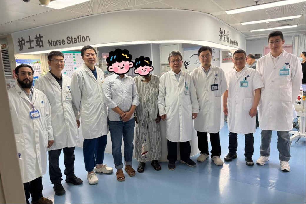
Recently, the team led by Geng Zhimin from the Biliary Surgery Subspecialty of the Department of Hepatobiliary Surgery at the First Affiliated Hospital (FAH) of Xi’an Jiaotong University (XJTU) successfully performed laparoscopic radical resection of hilar cholangiocarcinoma on a 62-year-old Bangladeshi patient. The patient recovered, was discharged just eight days after surgery, and returned to Bangladesh.
The patient had developed symptoms of jaundice and pruritus since August 2025 and had undergone multiple ERCP procedures and stent placements at local hospitals. However, the condition repeatedly recurred without a definitive diagnosis. In March 2026, the FAH received a request for an international consultation. Based on the patient’s medical history, Geng Zhimin’s team strongly suspected hilar cholangiocarcinoma and recommended endoscopic biopsy during ERCP to establish a definitive diagnosis. On April 28, the patient was admitted to the FAH for comprehensive examinations. Following discussion by the multidisciplinary team (MDT) for biliary tumors and confirmation by biopsy, the patient was diagnosed with Bismuth type IIIb hilar cholangiocarcinoma.
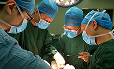
On May 11, under the guidance of Chief Physician Geng Zhimin, Associate Chief Physician Zhang Dong and Attending Physicians Chen Chen and Lei Jianjun performed laparoscopic radical resection of hilar cholangiocarcinoma combined with left hemihepatectomy, successfully achieving R0 resection. Several advanced technologies played key roles throughout the diagnosis and treatment process. Before surgery, ERCP combined with peroral cholangioscopy was used for precise endoscopic mapping to determine the nature of the lesion and the extent of tumor invasion, providing a basis for surgical planning. Imaging examinations and three-dimensional liver reconstruction were used to precisely calculate the future liver remnant volume, while the MDT conducted a comprehensive assessment to rule out the risk of tumor metastasis, providing robust safeguards for surgical safety.
This was a highly complex minimally invasive procedure involving left hemihepatectomy, caudate lobectomy, and bile duct reconstruction. Postoperative pathological examination showed negative resection margins. The patient developed no complications and recovered well.
In view of the patient’s status as an international patient, the team developed postoperative treatment and follow-up plans in accordance with international standards and established a dedicated communication channel, providing full-cycle management from admission, surgery, and rehabilitation to follow-up after the patient returned to his home country. The successful treatment of this complex cross-border case fully demonstrated the comprehensive strengths of the FAH’s Biliary Surgery Subspecialty in minimally invasive techniques, endoscopic diagnosis and treatment, and multidisciplinary collaboration.