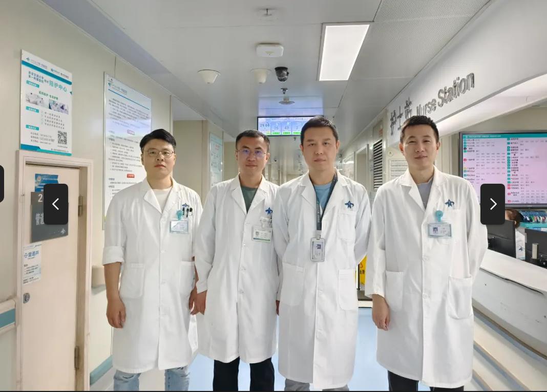
Recently, Associate Professor Li Qi’s team from Department of Neurosurgery of the First Affiliated Hospital (FAH) of Xi'an Jiaotong University (XJTU) performed intracranial tumor resection via fronto-basal interhemispheric approach during craniotomy under microscope combined with the latest 4K3D neuroendoscope for a 12-year-old child with craniopharyngioma. According to authoritative literature review, it is the first case of this kind worldwide.
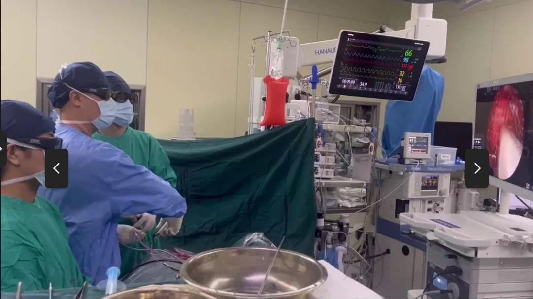
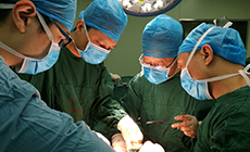
A 12-year-old patient was diagnosed with a space-occupying lesion in the sellar region in a local hospital. The patient was subsequently admitted to Department of Neurosurgery of FAH, Associate Chief Physician Li Qi considered the possibility of craniopharyngioma and growth retardation. Due to excessively narrow nasal cavity and lack of paranasal sinus pneumatization, it is impossible to perform nasal surgery. After a team discussion led by Li Qi and a general consultation led by Director Zhang Hua, it was determined to adopt intracranial tumor resection via fronto-basal interhemispheric approach during craniotomy under microscope combined with the latest 4K3D neuroendoscope. Intraoperatively, bilateral frontal lobes were carefully separated by dynamic traction technology under the microscope, and it was found that the tumor had been compressed to the optic chiasma. The tumor located on the suprasellar part was carefully resected to relieve the compression of the optic chiasma. Because the interior of the saddle was a blind area under the microscope, Li Qi adopted the most advanced 4K3D neuroendoscope worldwide to insert the detector into the blind area through the surgical channel, and found that a large quantity of tumors remained in the saddle and adhered closely to the pituitary gland and the inner wall of cavernous sinus. Then, he utilized angled neuroendoscopic surgical instruments to complete total resection of residual tumors in the sella, protect pituitary and cavernous sinus from damage, completely remove the craniopharyngioma, and minimize the risk of postoperative recurrence.
Skull base tumor surgery is highly challenging in the field of neurosurgery. At present, endoscopic sinus surgery is recognized as the surgical treatment for sellar lesions, especially pituitary tumors. The images of 4K3D neuroendoscope yield high stereoscopic sense, high resolution, increased image details, and a more thorough and accurate understanding of the spatial relationship, which is beneficial to identify the neurovascular structure in the dura mater during tumor resection, improve the depth perception and hand-eye coordination, enhance the safety and efficacy of surgery, alleviate postoperative complications, and play a critical role in the resection of sellar tumors in the field of neurosurgery.