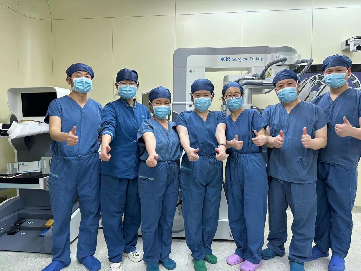
On the afternoon of January 26, 2024, the team led by Professor Li Qiling, Deputy Director of Department of Obstetrics and Gynecology, successfully performed robotic single-port laparoscopic staged surgical resection of granulosa cell tumors in the East District of the First Affiliated Hospital (FAH) of Xi’an Jiaotong University (XJTU). According to literature review, this is the first case of robotic single-port laparoscopic resection of the whole uterus + adnexa + the greater omentum in China.
On January 10, 2024, a 40-year-old female patient underwent resection of the right adnexa via a longitudinal abdominal incision in Yulin No.2 Hospital due to a huge cystic mass in the right adnexa. Postoperative pathological results revealed "granulosa cell tumors complicated with cystic lesions in right ovarian". The patient hoped to receive secondary surgery, especially minimally invasive surgery. Hence, she was admitted to the FAH on January 24, 2024.
Pathological examination after right oophorectomy confirmed granulosa cell tumors of the ovary. She had no need for fertility. Comprehensive staged surgery for ovarian tumors should be completed, involving the whole uterus, left adnexa and the greater omentectomy. The patient had undergone three open surgeries. Physical examination upon admission showed multiple surgical incisions in the lower abdomen. At postoperative 2 weeks, tissue edema and adhesion could be seen, all of which made it difficult to conduct comprehensive re-staged surgery for ovarian tumors. Considering pathological results and her desire to receive minimally invasive surgery as soon as possible, Professor Li Qiling's team decided to adopt an innovative treatment protocol after meticulous evaluation, adopting domestic independently designed and manufactured single-port robot-assisted laparoscopic re-staged surgery for ovarian tumors.

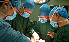
On January 26, after careful preoperative preparation, a 2.0-cm umbilical incision was created by Professor Li Qiling as chief surgeon, with the cooperation of Associate Professor Xue Yan and Attending Physician Wang Shuyuan, which successfully avoided the scars of previous surgical incisions. Intraoperatively, the patient's position was reasonably designed. Without other auxiliary ports, robotic single-port laparoscopic resection of the whole uterus, the left adnexa and the greater omentum, and the release of pelvic and abdominal adhesion were successfully completed. The operation time was 3.5 h and intraoperative blood loss was 50 mL. The patient was discharged successfully at postoperative 4 d. This procedure maximized the minimally invasive concept, leaving minimal internal and external trauma and almost no surgical scars on the body surface after surgery. The patient was rapidly recovered and the length of hospital stay was shortened, demonstrating the concept of "enhanced recovery after surgery".
This is the first case of robotic single-port laparoscopic resection of the whole uterus, the left adnexa and the greater omentum via umbilical approach, and the release of pelvic and abdominal adhesion in China. The snake-shaped operating arm of the single-port robot integrates both force load and flexibility, which can not only pull and move, but also bend around in all directions. The surgeons can accurately and stably complete each surgical procedure in narrow space.